Lesson 1,
Topic 1
In Progress
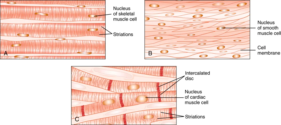
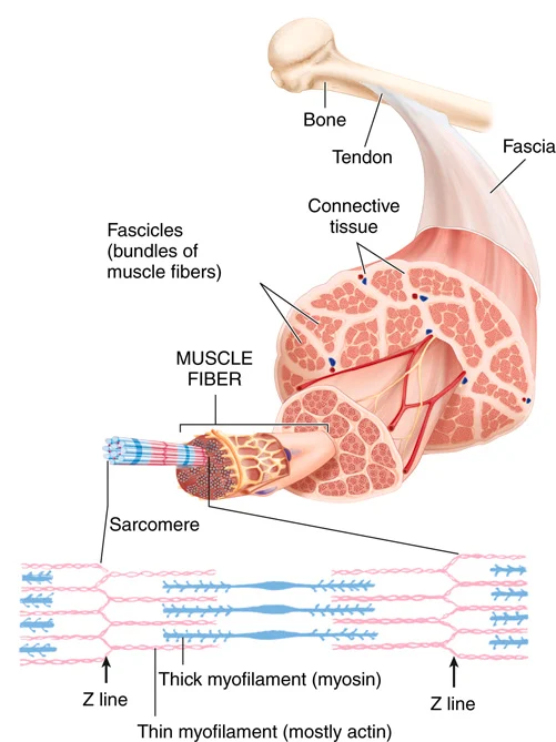
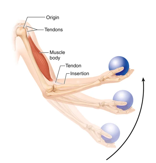
Anatomy of the Muscular System
| Movement | Definition or Example |
|---|---|
Flexion | Reduces the angle of the joint and brings the two bones closer together |
Extension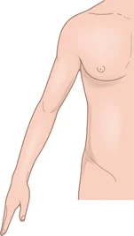 | The opposite of flexion; increases the angle or distance between two bones or parts of the body |
Hyperextension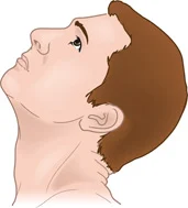 | Extension 180 degrees (e.g., the neck is extended backward or the toes are pointed downward) |
Abduction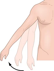 | Moving the body part away from the midline or median plane of the body |
Adduction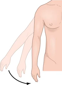 | The opposite of abduction; moving the body part toward the midline of the body |
Rotation | Moving a bone around its central axis; common in ball-and-socket joints |
Circumduction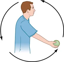 | Circular movement of a limb; a combination of abduction, adduction, extension, and flexion |
Dorsiflexion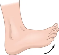 | Moving the instep of the foot up and dorsally, reducing the angle between the foot and the leg |
| Table Continued |
| Movement | Definition or Example |
|---|---|
Plantar flexion | A toe-down movement of the foot at the ankle; increases the angle of the joint |
Eversion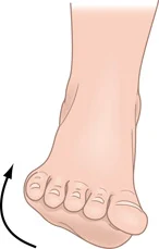 | Turning the sole of the foot laterally, or outward |
Inversion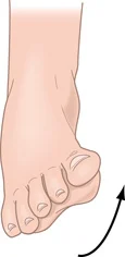 | The opposite of eversion; turning the sole of the foot medially, or inward |
Pronation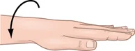 | Rotation of the forearm that turns the palm of the hand downward, or posteriorly |
Supination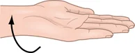 | The opposite of pronation; rotation of the forearm that turns the palm of the hand upward, or anteriorly |