Cells of the Nervous System
April 11, 2024
Cells of the Nervous System
Learning Objective: Differentiate among the cells of the nervous system.
The nervous system is made up of two types of cells:
• Neurons: Also called parenchymal cells, which carry out the work of the nervous system.
• Neuroglia: Also called stromal cells or glia. These cells provide a supportive function for the neurons.
Neurons
Learning Objective: Discuss anatomy of neurons.
The nervous system is composed of nerve cells, or neurons. Neurons are composed of a cell body, dendrites, and an axon. The dendrites are short projections from the cell body. They pick up electrical impulses, often from other neurons, and send the signals to the cell body. The impulse is then passed to the axon, which is a long extension off the cell body. Some axons can be over 3 feet long. The axon transmits the impulse to other dendrites, glands, or muscles.
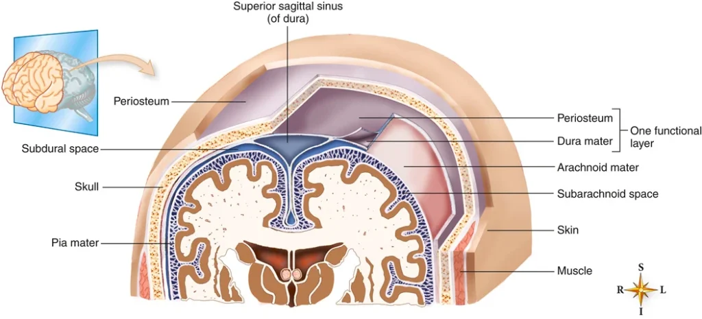
FIGURE 22.4 Protective coverings of the brain. Modified from Patton K, Thibodeau G: Anatomy and physiology, ed 9, St. Louis, 2016, Mosby.
Table 26.5
Cranial Nerves
| Number | Name | Function | How it is Tested |
|---|---|---|---|
| I | Olfactory | Sensory function: Smell | Identifying familiar odors while the eyes are closed. |
| II | Optic | Sensory function: Vision | A visual acuity test may be given. A light may be shined in the eyes. |
| III | Oculomotor | Sensory and motor functions: Eye movement, pupil constriction and accommodation | The pupil is examined using a light. The patient may be asked to follow the light with the eyes without moving the head. |
| IV | Trochlear | Motor function: Eye movement | Testing to see if the eyes can follow a moving light, as described for cranial nerve III. |
| V | Trigeminal | Sensory and motor functions: Muscles for chewing, general sensations from the anterior half of the head, including the face and meninges | The provider may feel the face in different locations and have the patient clench the teeth. |
| VI | Abducens | Motor function: Eye movement | Testing to see if the eyes can follow a moving light, as described for cranial nerve III. |
| VII | Facial | Sensory and motor functions: Muscles used for facial expressions; tearing, salivation, and taste | Patient may be asked to identify common tastes, puff cheeks, smile, wrinkle forehead, and close eyes tightly. |
| VIII | Vestibulocochlear | Sensory function: Hearing and equilibrium | Assessed using a hearing acuity test. |
| IX | Glossopharyngeal | Sensory and motor functions: Swallowing and taste | Gag reflex is tested with a tongue blade. |
| X | Vagus | Sensory and motor functions: Breathing, speech, sweating, regulating heartbeat, stimulating muscles of the gastric region | Patient is asked to say “ahh,” swallow, and talk (voice quality assessed). Gag reflex is tested with a tongue blade. |
| XI | Spinal accessory | Motor function: Shoulder and head movements | Assessed by turning the head from side to side against mild resistance and shrugging the shoulders. |
| XII | Hypoglossal | Motor function: Tongue movements | Assessed by sticking out and moving the tongue from side to side. |
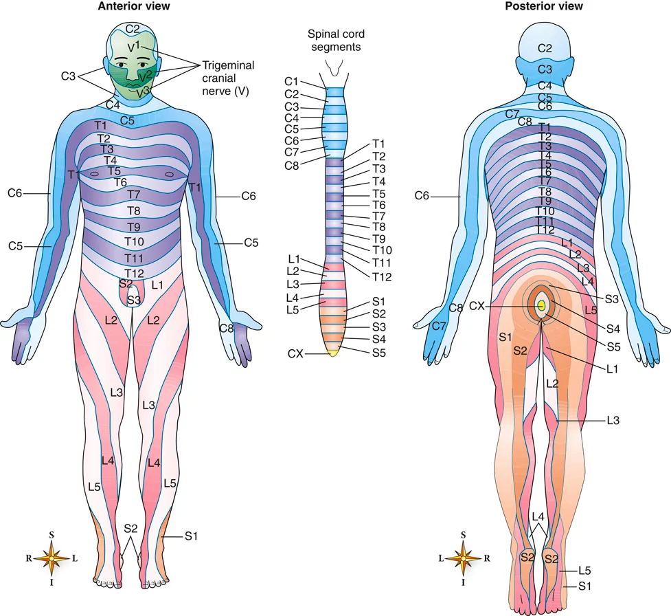
FIGURE 22.5 Map of the dermatomes. From Patton K, Thibodeau G: The human body in health & disease, ed 7, St. Louis, 2018, Mosby.
Neuroglia
Learning Objective: Describe the different types of neuroglia cells.
Neuroglial cells care for and support neurons throughout the body. These specialized cells perform specific functions within the nervous system. There are several types of neuroglia, including the following:
• Schwann cells: Form the myelin sheath, which covers the axons of peripheral nerves.
• Astrocytes: Help form the blood-brain barrier (BBB), which closely regulates what substances enter the brain tissue. Oxygen, water, and glucose molecules easily pass into the brain. Many chemicals and drugs are prevented from moving into brain tissue.
• Microglia: Found in the CNS. If brain tissue is inflamed or damaged, microglia engulf and destroy microorganisms or debris.
• Oligodendrocytes: Found in the CNS; they myelinate the CNS axons and help hold nerve fibers together.
22.3
Critical Thinking Application
What type of cell produces the myelin sheath that surrounds some nerve axons?