Central Nervous System
April 11, 2024
Central Nervous System
Learning Objective: Differentiate among the structures of the central nervous system.
The central nervous system is composed of the brain and spinal cord. The following sections examine the brain and spinal cord.
Brain
Learning Objective: Describe the anatomy of the brain.
The brain, enclosed in the skull, is one of the most complex organs of the body. It is divided into the cerebrum, cerebellum, diencephalon, and brainstem (FIGURE 22.1).
Cerebrum
The cerebrum is the largest portion of the brain and is divided into two hemispheres by a deep fissure. The right hemisphere controls artistic functions, such as drawing, rhythm, and picture memory. The left hemisphere controls verbal functions, such as reading, writing, speaking, and mathematic calculations. The hemispheres are connected by the corpus callosum, a bundle of nerve tissue that facilitates communication between the two sides of the brain. If the corpus callosum only partially develops or is absent, the person is diagnosed with corpus callosum agenesis.
Most of the signals between the brain and body cross over. This means that the left cerebral hemisphere primarily controls the right side of the body, and the right hemisphere primarily controls the left side of the body. For instance, if a person has a stroke affecting the right hemisphere, the symptoms will be seen on the left side of the body.
The surfaces of the hemispheres are covered by gray matter, or the cerebral cortex. The cerebral cortex is a thin layer ranging from 1 to 4.5 mm in thickness. Most of the information processing in the brain takes place in the cerebral cortex. Gyri add more surface area, thus increasing the amount of information that can be processed. Sulci are also visible in the cerebral cortex.
Lobes
Each cerebral hemisphere is further divided into four sections, called lobes. These are the functions of the lobes:
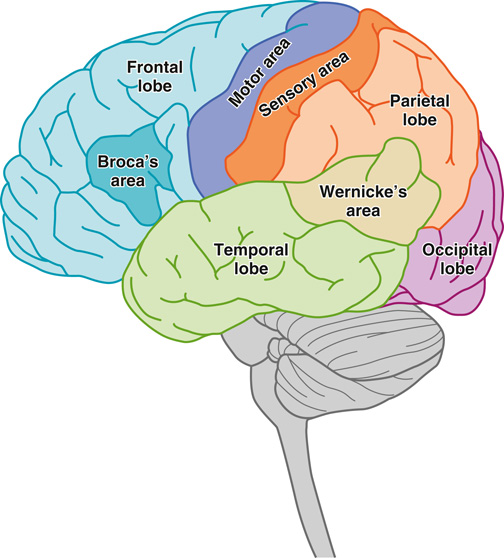
FIGURE 22.2 The lobes of the cerebrum.
The diencephalon is located just above the brainstem and is almost surrounded by the cerebral hemispheres. It serves as a relay station for sensory input neurons and other parts of the brain. It plays an important role in the functioning of the CNS, endocrine system, and limbic system. The diencephalon is composed of the posterior pituitary gland and the pineal gland, which are part of the endocrine system. The diencephalon also includes two other parts:
Cerebellum
The cerebellum is located below the occipital lobe of the cerebrum. It is also covered by the cortex. The cerebellum gathers input from other parts of the brain and spinal cord to provide accurate timing for coordinated smooth movements. The cerebellum coordinates the equilibrium or balance, posture, and muscle coordination. Damage to the cerebellum caused by a stroke can cause nausea, dizziness, and balance and coordination issues.
Diencephalon
The diencephalon is located just above the brainstem and is almost surrounded by the cerebral hemispheres. It serves as a relay station for sensory input neurons and other parts of the brain. It plays an important role in the functioning of the CNS, endocrine system, and limbic system. The diencephalon is composed of the posterior pituitary gland and the pineal gland, which are part of the endocrine system. The diencephalon also includes two other parts:
• Thalamus: Processes information going to and from the body and the cerebrum.
• Hypothalamus: Controls body temperature, hunger, and thirst; part of the limbic system.
A tract of nerve cells connects the thalamus and hypothalamus to the hippocampus. The hippocampus works with memories, sending memories to cerebral hemispheres for storage (long-term memories) and then retrieving them when needed.
22.1
Critical Thinking Application
Nancy has roomed Elaine. While Nancy is taking her medical history, Elaine mentions that she feels unsteady from time to time and loses her balance a few times. What part of the brain is involved in maintaining balance?
Brainstem
The brainstem connects the cerebral hemispheres to the spinal cord. It controls the flow of information from the body to the brain. The brainstem is composed of these structures:
• Midbrain: Connects the hemispheres of the cerebrum with the pons. It serves as a relay center for auditory, visual, and motor information. It also contains centers to regulate pupillary reflexes and eye movements.
• Pons: Serves as a bridge between the medulla oblongata and the cerebrum and helps regulate respiration.
• Medulla oblongata: Lowest part of the brainstem. It contains vital centers of life (e.g., cardiac, respiratory, and vasomotor centers) that regulate the heart rate, respiration, and the diameter of blood vessels, which affect the blood pressure. Nonvital centers also are found in the medulla oblongata, including centers that regulate coughing, sneezing, hiccupping, vomiting, and swallowing. Some medications work to quiet these centers.
Spinal Cord
Learning Objective: Describe the anatomy of the spinal cord.
The spinal cord is a bundle of nervous tissue. It extends from the medulla oblongata to about the second lumbar vertebra (FIGURE 22.3). The spinal cord passes through the foramen magnum in the skull and is protected by the vertebrae. It is covered by meninges. The spinal cord is composed of the cell bodies of motor neurons (gray matter) and the myelin-covered axons. Thirty-one pairs of spinal nerves extend from the spinal cord. Each nerve stimulates a specific organ or area of the body. The spinal cord carries messages between the spinal nerves and the brain. This can slow with age.
Meninges
Meninges are a protective covering around the brain and spinal cord. They are composed of three membranes (FIGURE 22.4):
• Dura mater: Outer layer of meninges; made up of a tough white fibrous connective tissue. The space below the dura mater is called the subdural space, which contains tiny blood vessels.
• Arachnoid mater: Middle layer of meninges; made up of a thin layer of threadlike strands resembling a cobweb. The space below the arachnoid is called the subarachnoid space, which is filled with cerebrospinal fluid (CSF) and blood vessels.
• Pia mater: Innermost layer of meninges; a thin, highly vascular membrane that is tightly bound to the surface of the spinal cord and brain.
The ventricular system in the brain is made up of four connected ventricles (spaces or cavities). CSF originates in the choroid plexus in the ventricles. CSF is a clear fluid that resembles water, and it has three roles:
• Cushions the brain and spinal cord
• Removes waste products from cerebral metabolism
• Supplies nutrients to the nervous system tissues
CSF flows through the ventricles, exits into the cisterns at the base of the brain, and moves around the brain and spinal cord. Eventually, CSF is reabsorbed into the bloodstream. The balance between CSFs production and absorption is critical. Excessive amounts of CSF result in an abnormal widening of the ventricles, which can put pressure on the brain tissue and lead to hydrocephalus .