Human Chorionic Gonadotropin Levels
June 23, 2023
Human Chorionic Gonadotropin Levels
HCG is the hormone assessed in pregnancy tests. This hormone can be detected in both urine and blood.
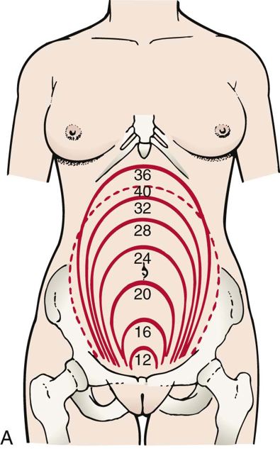
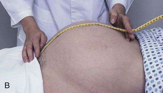
Box 25-2 Factors That Influence Fundal Height Measurement
- • Maternal position
- • Maternal stature
- • Maternal obesity
- • Presence of uterine fibroids
- • Elevations in amniotic fluid volume
Data from Davidson M, London M, Ladewig P: Olds’ maternal-newborn nursing and women’s health across the lifespan, ed 9, Upper Saddle River, N.J., 2010, Pearson.
Testing for HCG can be quantitative or qualitative. Quantitative testing considers the level or amount of the hormone in the specimen. Quantitative results are obtained from a blood specimen. Qualitative testing simply determines the presence or absence of a substance. Qualitative testing for HCG may be performed on both the blood and the urine. During pregnancy, the levels of HCG may be used to assess the viability and gestation of the pregnancy. HCG levels that are less than anticipated for a specific point in gestation may signal a pregnancy that is no longer viable. Quantitative HCG levels may be reported every few days to determine whether they are rising as in a normally developing pregnancy or whether they are declining in a manner consistent with miscarriage.
Ultrasound Scan
During ultrasonography, high-frequency sound waves are used to visualize the fetus. Because soft tissue is visualized, this noninvasive tool can be used to determine gestational age, monitor fetal growth, determine the number of fetuses and the location of the placenta, estimate the volume of amniotic fluid, and detect anomalies. It can also be used in conjunction with invasive tests such as amniocentesis. The gestational age often determines the type of ultrasound scan performed. When used to determine the gestational age, the health care provider performs a series of measurements. In the first trimester, the fetal crown-rump length is used. Crown-rump length is the measurement from the top of the fetal head to the buttocks. This recording is correct within 2 to 5 days. As the pregnancy progresses and the fetal positioning changes, this becomes an ineffective means to measure gestational age. In the second trimester, the examiner measures fetal head circumference, biparietal diameter, and femur length to calculate gestational age. The degree of accuracy is 4 to 9 days. Estimation of fetal age in the third trimester is even less accurate because of a variety of interrelated factors that include genetics, gender differences, and maternal-related concerns (Behrman and Butler, 2007). During the first trimester, the transvaginal ultrasound scan may be used. The small distance between the probe, which is inserted vaginally, and the uterus make the tranvaginal ultrasound scan a prime tool for assessment of the early pregnancy (Gabbe, 2012) to confirm the presence and viability of the pregnancy.
After the first trimester, the abdominal ultrasound scan is preferred. It may be used singularly or in conjunction with other methods of maternal fetal assessment. Early in pregnancy, the mother is required to drink a quart of water and not void. This allows for the bladder to elevate the uterus for better viewing with the technology.
A growing number of nurses perform ultrasound scans and biophysical profiles (BPPs; a system of estimating current fetal status by analyzing five variables via ultrasound scan and nonstress testing). However, most nurses are primarily involved in counseling and educating women about the procedure (see Patient Teaching box on ultrasound scan examination).
Maternal Serum Alpha-Fetoprotein Screening (Quadruple Marker Screening)
Maternal serum alpha-fetoprotein screening is commonly referred to as the Quadruple Marker Screening. Note that the assessment is a screening with results that provide a prediction for the occurrence of certain
birth defects. The results do not provide a definitive diagnosis or presence of a disorder. Screening of the levels may be performed to identify the presence of chromosomal abnormalities, such as Down syndrome, or birth defects, such as neural tube defects. The factors assessed with the screening are alpha fetoprotein, estriol levels, HCG, and inhibin A. Alpha-fetoprotein is a protein produced by the fetal yoke sac. Serum testing should be done between 16 and 18 weeks of gestation (Rakel, 2011). Elevated levels are suggestive of neural tube defects, whereas low levels may suggest a fetus with Down syndrome (trisomy 21). Screening results may be influenced by maternal age and weight, gestational age, and the presence of more than one fetus.
Abnormal findings result in additional testing. Ultrasound scan may be performed to assess fetal